My favorite app
Great app, easy to use. Helps our small business save time and money to create different marketing materials professionally. Thank you.
Begin by uploading your de-identified MRI scan. The tool supports standard image formats like PNG and JPG.
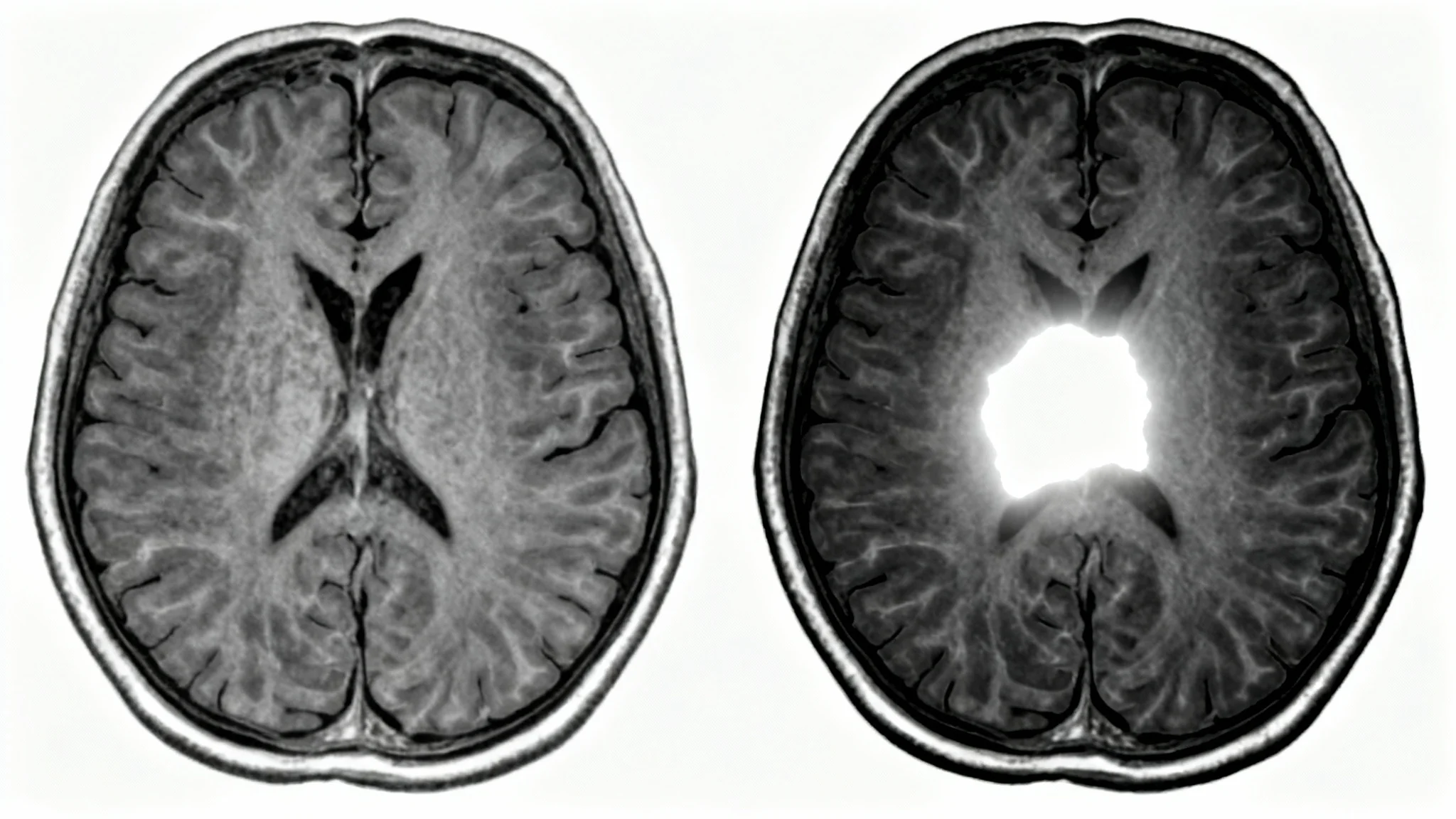
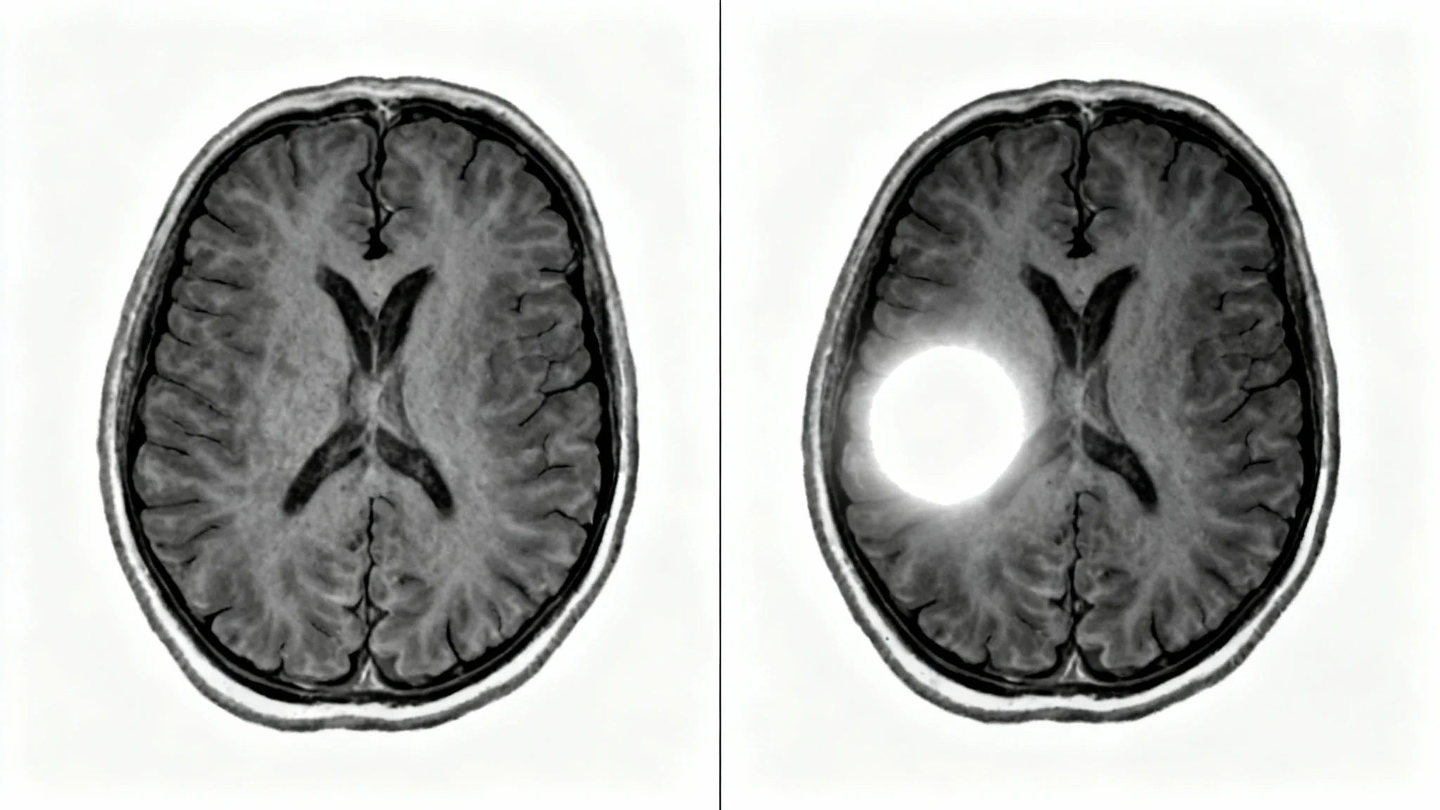
Provide a simple text instruction describing what you want to enhance. For example, 'Sharpen the details of the post-contrast T1 brain scan'.
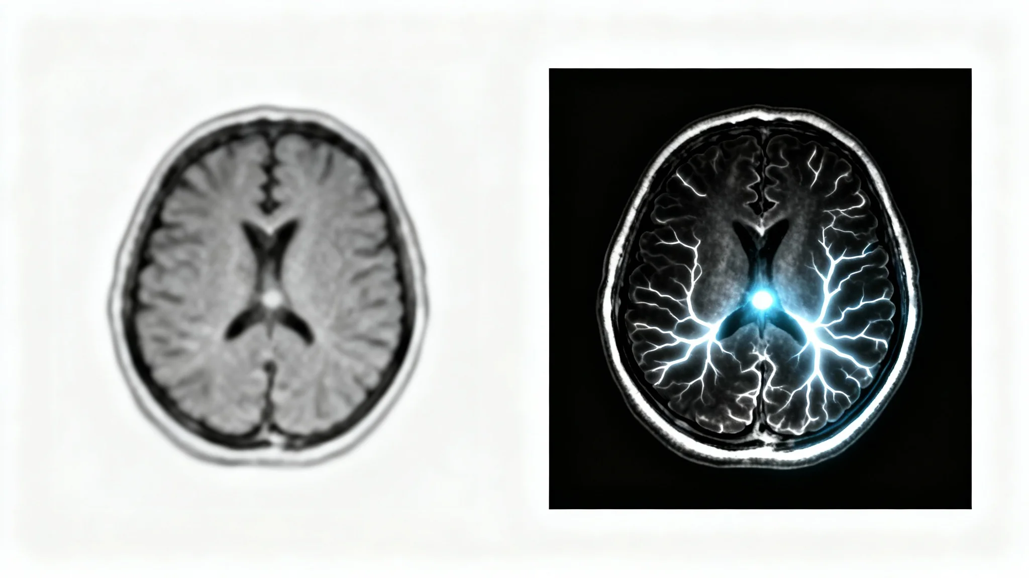
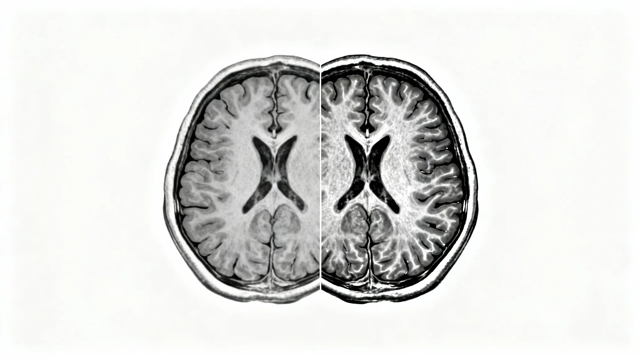
The AI will analyze your scan and prompt to generate a new image with improved contrast and clarity in just a few seconds.
Your high-resolution, enhanced MRI image is ready for download. Use it for presentations, research, or to support your analysis.
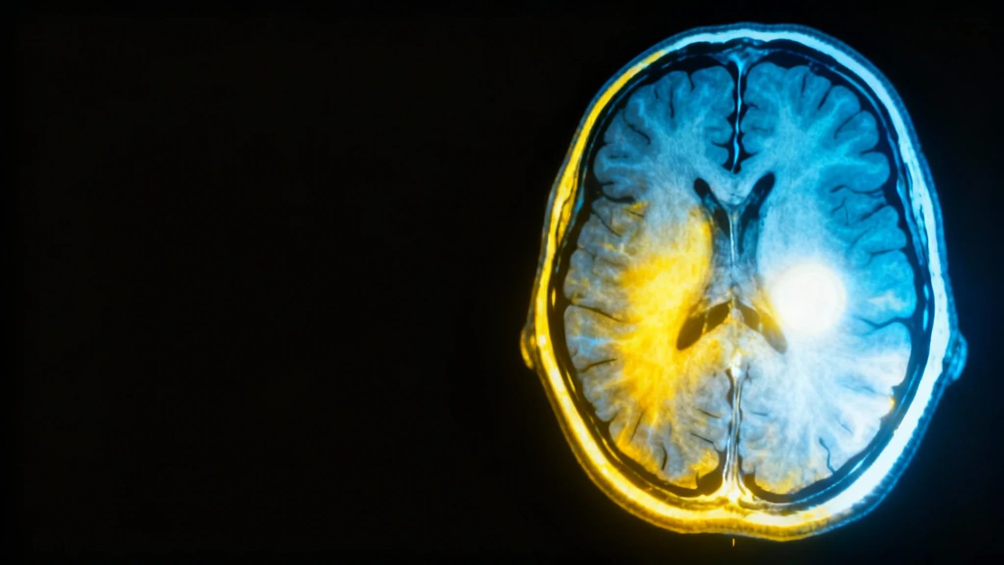
AI MRI contrast enhancement is a process where a deep learning algorithm analyzes an MRI scan and computationally improves its contrast. This can help make anatomical structures and pathological findings clearer and easier to interpret without altering the original scan data.
To use the tool, you first upload an MRI image in a standard format like PNG or JPG. Then, you can write a text prompt specifying your enhancement needs, such as 'enhance contrast to show lesion boundaries.' The AI will then process the image and provide an enhanced version for you to download.
This tool is designed for post-processing and enhancing the contrast of existing images. While some research explores using AI to simulate contrast or enhance low-dose contrast scans, this tool enhances scans that have already been taken. It should not be considered a substitute for gadolinium-based contrast agents in clinical practice.
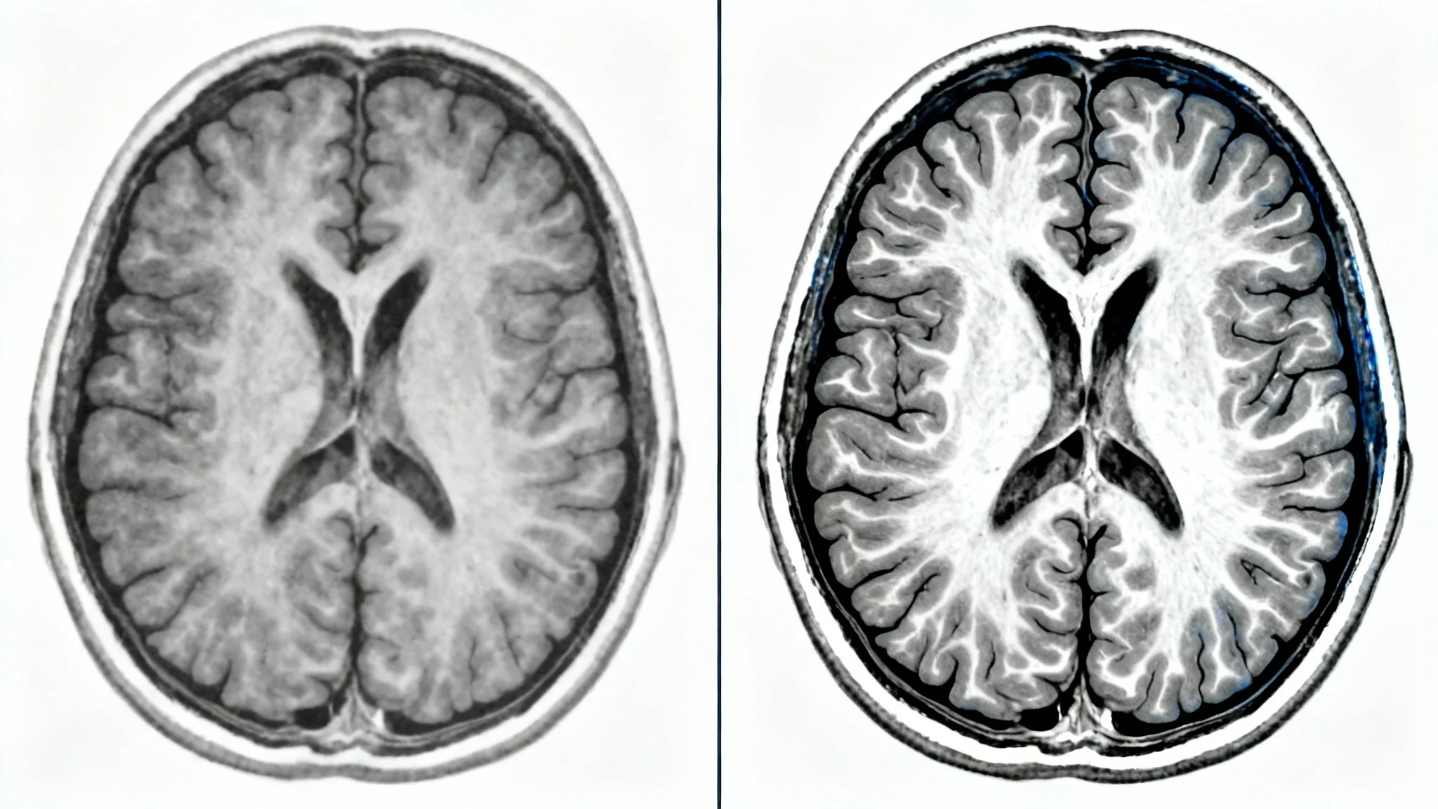
The tool is versatile and can be used to enhance a variety of MRI sequences, such as T1-weighted, T2-weighted, and FLAIR images across different body parts, including brain, joint, and body scans. The goal is to improve the visual quality of any uploaded MRI scan.
Yes, patient privacy is a priority. All uploaded data is handled in a secure environment. However, it is recommended to de-identify all images by removing any patient-identifying information before uploading them to any online tool.
This AI tool is designed to assist medical professionals and researchers by enhancing image quality for better visualization. It is not a certified medical device and should not be used as a primary tool for clinical diagnosis. All diagnostic decisions should be made by qualified medical professionals.
Great app, easy to use. Helps our small business save time and money to create different marketing materials professionally. Thank you.
I need a good background remover for my work and I've been through them all it feels like. This one is the best by far. It's so easy to use and the results always look amazing. Thank you Pixelcut!
It's amazingly easy and gives the perfect results for my bags and accessories. It looks like it was done by a professional photographer.